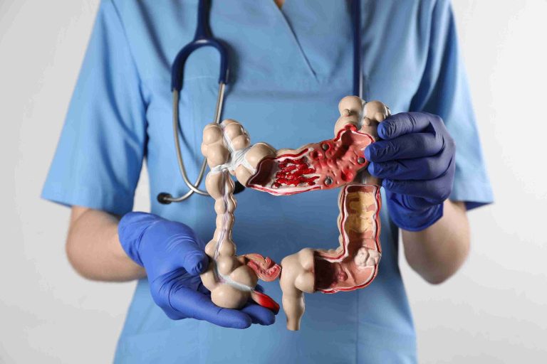
Esophageal varices are abnormal, enlarged veins in the esophagus, typically resulting from liver disease and increased pressure in the portal vein. Left untreated, esophageal varices can lead to life-threatening bleeding.
Gastroscopy plays a crucial role in diagnosing and treating this condition. This article will explore the significance of gastroscopy in managing esophageal varices and why it is considered a life-saving procedure for those at risk of variceal bleeding.
Understanding Esophageal Varices
Esophageal varices develop primarily in individuals with advanced liver disease, particularly cirrhosis. Cirrhosis scars the liver, impeding blood flow and increasing pressure in the portal vein (portal hypertension). As the pressure builds, blood is rerouted through smaller vessels, including those in the esophagus, which can swell and form varices.
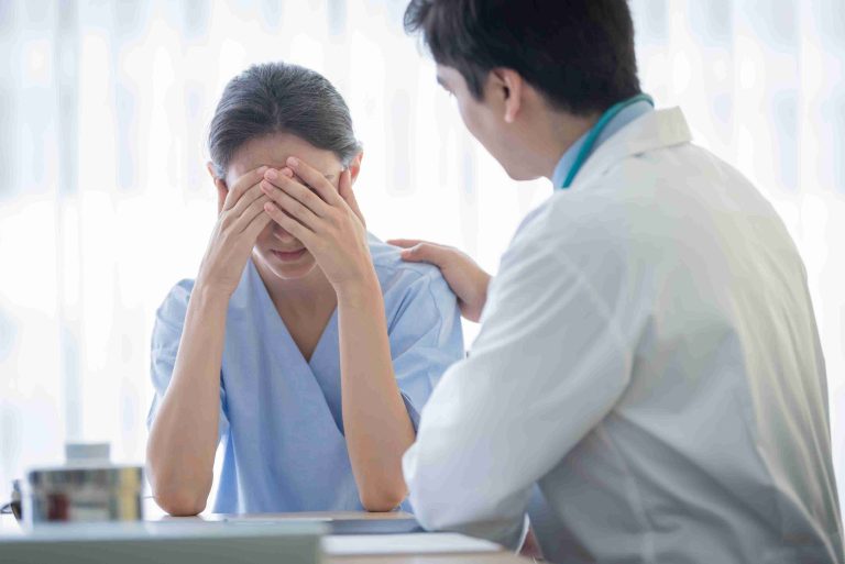
Although varices may not cause symptoms initially, they pose a significant risk because of their fragility. When these veins rupture, severe internal bleeding can occur, which can be fatal if not promptly treated. Early detection and timely management are crucial for individuals with esophageal varices, and this is where gastroscopy becomes vital.
Gastroscopy: A Diagnostic and Therapeutic Tool
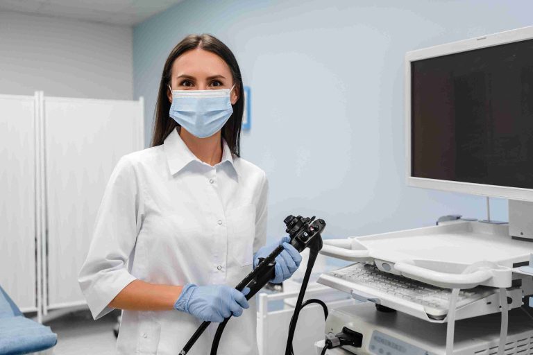
Gastroscopy is a procedure where a thin, flexible tube equipped with a light and camera (endoscope) is inserted through the mouth to examine the esophagus, stomach, and upper part of the small intestine. This minimally invasive procedure allows doctors to not only diagnose esophageal varices but also treat them during the same session if needed.
In the case of esophageal varices, gastroscopy is used to visualize the size, number, and appearance of the varices, determining their risk of bleeding. Physicians can assess whether the varices are likely to rupture and intervene if necessary. This early intervention can significantly reduce the risk of a life-threatening event, making gastroscopy an essential tool in managing the condition.
How Gastroscopy Treats Esophageal Varices
One of the primary therapeutic uses of gastroscopy in esophageal varices is endoscopic band ligation. This procedure involves placing rubber bands around the varices, cutting off their blood supply and causing them to shrink. This process is particularly effective in preventing variceal bleeding or re-bleeding in patients who have already experienced a rupture.
Endoscopic sclerotherapy is another treatment method where a solution is injected into the varices to make them shrink and close off. While not as commonly used as band ligation due to potential complications, sclerotherapy can be effective in certain situations where banding may not be feasible.
Both treatments are performed during gastroscopy, and the decision between ligation or sclerotherapy depends on the size and severity of the varices, the patient’s overall health, and whether bleeding has already occurred.
Monitoring and Preventing Re-Bleeding
Gastroscopy plays a central role not just in treating active varices but also in monitoring patients for recurrence. After an initial treatment, patients often require follow-up gastroscopies to check if new varices have formed or if existing ones have returned. Regular monitoring through gastroscopy allows for timely intervention, significantly reducing the risk of a major bleeding event.
In high-risk patients, prophylactic band ligation can also be performed to prevent the development of esophageal varices before they become a critical issue. By closely following at-risk patients with gastroscopy, doctors can prevent complications from worsening, contributing to better long-term outcomes.
Why Early Detection Through Gastroscopy is Vital
Early detection of esophageal varices through gastroscopy is crucial because varices are often asymptomatic until a rupture occurs. Once bleeding starts, the patient is at significant risk, and emergency treatment becomes more challenging and carries higher risks. Through early gastroscopic screening, particularly in patients with cirrhosis or chronic liver disease, doctors can identify varices before they become dangerous.
In individuals with liver disease, a proactive approach to screening through gastroscopy is essential for reducing the risk of bleeding. Studies have shown that regular surveillance of high-risk individuals significantly improves survival rates, as treatment can be administered before a rupture occurs. The procedure itself is relatively safe, with minimal complications, making it an excellent tool for managing esophageal varices.
Potential Risks and Complications of Gastroscopy
While gastroscopy is generally a safe procedure, like any medical intervention, it carries some risks. Patients may experience discomfort or a sore throat following the procedure. In rare cases, there may be a risk of perforation of the esophagus, bleeding, or infection, particularly if a therapeutic procedure such as banding or sclerotherapy is performed. However, the benefits of gastroscopy in preventing life-threatening variceal bleeding far outweigh the risks.
To minimize potential complications, the procedure is often performed under sedation, ensuring the patient is relaxed and comfortable throughout. The recovery time is usually short, with most patients able to resume normal activities within a day.
Conclusion
Gastroscopy is an invaluable tool in both diagnosing and treating esophageal varices. It allows doctors to assess the condition of the varices, perform immediate therapeutic interventions like banding or sclerotherapy, and monitor the patient for recurrence.
For individuals with liver disease, early gastroscopy screenings can prevent life-threatening complications by addressing varices before they bleed. Given the risks associated with esophageal varices, particularly in those with cirrhosis, regular gastroscopic evaluation is essential for effective management and better long-term outcomes.